Diagnostic Services Available at Our Clinic in Thornleigh, NSW
Ultrasound Scan
When symptoms involve abdominal organs or soft tissue, doctors often suggest ultrasound imaging. The scan uses sound waves to produce real-time images of organs, blood vessels, and surrounding structures. During the scan, expert sonographers capture images while observing movement within the body.
Doctors often rely on ultrasound to investigate issues including:
- Conditions involving the liver, kidneys, gallbladder, or pancreas
- Pregnancy development and assessing foetal growth
- Cysts, fluid collections, or soft tissue abnormalities
- Blood circulation through arteries and veins
Ultrasound often provides doctors with a clear starting point for investigating a wide range of symptoms.
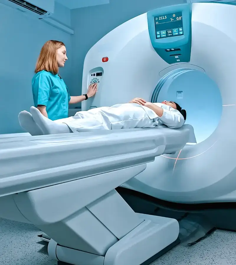
CT Scan (Computed Tomography)
When doctors require deeper internal detail, CT (Computed tomography) imaging often provides the next level of diagnostic clarity. It creates cross-sectional images using which doctors can examine organs, bones, blood vessels, and complex injuries.
Modern CT scanners capture multiple image layers within seconds while maintaining radiation protocols consistent with Australian medical standards.
CT imaging plays an important role in identifying conditions such as:
- Internal bleeding following trauma or accidents
- Tumours, infections, or abnormal growths in organs
- Lung disease, chest infections, or pulmonary conditions
- Complex bone fractures and spinal injuries
These images provide doctors with the detailed view required to confirm complex diagnoses.
X-Ray Scan
For many injuries and chest conditions, doctors begin investigations with standard X-ray imaging. It remains one of the most widely used diagnostic tools. The scan produces clear images of bones and certain tissues, allowing clinicians to assess injuries quickly.
Digital X-ray systems capture images with improved clarity while allowing radiographers to adjust positioning carefully according to the referral request. It helps clinicians:
- Detect bone fractures, dislocations, and joint abnormalities
- Identify lung infections such as pneumonia or other chest conditions
- Monitor arthritis and degenerative joint changes
- Assess bone alignment after injury or orthopaedic treatment
X-ray imaging often provides fast answers for many common medical concerns.
Cortisone Injections
Image-guided cortisone injections assist patients experiencing joint inflammation, tendon irritation, or persistent musculoskeletal pain. Orthopaedic specialists and sports physicians often request these procedures when targeted treatment becomes necessary.
During the procedure, imaging guidance allows medication to be placed directly into the affected joint or soft tissue structure.
These injections are commonly used to manage conditions such as:
- Inflammation within shoulder, hip, knee, or spinal joints
- Bursitis and tendon irritation caused by overuse injuries
- Arthritis
- Degenerative joint disease
Cortisone injections also support rehabilitation for persistent sports-related conditions.
Often, these form a crucial part of a broader treatment plan recommended by the patient’s doctor.
Sports Imaging
Sports persons, whether beginners or professionals are prone to injuries during play. Since Thornleigh has a strong population of athletes, runners, and recreational players, our clinic provides high-quality imaging solutions to understand the sport-related problems.
These are commonly known as musculoskeletal imaging that allows doctors to examine structures such as cartilage, ligaments, tendons, and muscles in detail. These reveal:
- Ligament tears including ACL and ankle ligament injuries
- Tendon damage such as rotator cuff or Achilles injuries
- Muscle strains and soft tissue trauma
- Joint damage following impact or overuse
Post findings, doctors are able to guide rehabilitation programs or surgical planning, as per individual requirements.
Orthopantomogram (OPG)
Dentists request an OPG scan when they need a complete view of the teeth and jaw in a single image. The scan produces a panoramic X-ray of the mouth, including the teeth, jawbones, and surrounding structures.
Unlike small dental X-rays that focus on individual teeth, an OPG captures the full dental arch in one image. This wider perspective allows dentists to evaluate oral structures more thoroughly before treatment planning.
An OPG scan helps dentists assess dental structures and conditions such as:
- Impacted or unerupted wisdom teeth and monitoring their development
- Jawbone infections, cysts, tumours, and dental abscesses
- Jaw alignment and temporomandibular joint (TMJ) concerns
These are also beneficial when planning for orthodontic treatment, implants, or tooth extractions
DEXA Scan
Bone density imaging, commonly referred to as DEXA scan, plays an important role in monitoring long-term skeletal health. It measures bone mineral density in the spine and hip to assess bone strength and fracture risk.
Doctors utilise the DEXA scan results to:
- Diagnose osteoporosis and osteopenia before fractures occur
- Assess fracture risk in ageing adults
- Monitor bone health during long-term treatment
- Track bone density changes during osteoporosis management
The results help doctors guide preventative care and long-term treatment.

Parking
Three hour secure underground parking at Ed Square Town Centre
Public Transport
Edmondson Train Station Rd - 4 minute walk (300m)
Trusted Medical Imaging in Thornleigh
Medical imaging often sits at the centre of diagnosis. A GP may request an ultrasound for abdominal pain, while a sports physician might need CT imaging to understand joint damage. In these moments, reliable scans and precise reporting matter.
Our imaging centre provides carefully coordinated diagnostic services supported by experienced radiologists and qualified technologists. We use state-of-the-art scanners, monitoring devices, injector systems and other crucial medical equipment, all designed as per Australian radiation safety standards.
From the time you book medical imaging or other procedures with us, our experienced staff ensures you are well-informed throughout by following a few steps that include:
- Confirming referral details
- Explaining the scan before the examination begins
- Providing preparation guidance so patients arrive ready
- Positioning patients carefully
- Ensuring the scanner captures the correct area
- Reviewing images through a radiologist and sending reports to the referring doctor
This organised approach helps doctors receive the information they need while keeping the visit simple for patients.
Our Radiology Clinics Near You
Book Your Appointment Now
Choose a convenient location, and our team will guide you through preparation, timing, and referral requirements to keep things moving without unnecessary delays.
Frequently Asked Questions
Do I need to prepare for my scan before arriving? →
Preparation depends on the type of scan requested. Some ultrasound or CT scans may require fasting or drinking water beforehand, while X-rays usually require little preparation. Our team will be providing you with guidelines during the time of booking so you can come prepared, if need be.
Will the scan be uncomfortable or painful? →
Most imaging scans are quick and painless. You will need to remain still so the images are captured correctly. Some patients may find the process claustrophobic. In case, you happen to experience it, you can convey it with our team and they can guide you with a few breathing techniques that calm you during the process.
Are the facilities suitable for elderly patients or those with limited mobility? →
Yes. Our facility is wheelchair-accessible. Apart from that our staff will be continuously assisting if there is a requirement for additional support with positioning or moving safely during the examination.
What should I expect after a cortisone injection? →
Most patients resume normal activities shortly after the procedure. Mild soreness around the injection area may occur for a day or two. Your doctor will guide the next stage of treatment.